Home » Without Label » Cross Section Of A Bone : Structure and Function of the Musculoskeletal System ... - 100x first focus in the compact decalcified bone (cb) on the left part of the image, you can see small dots, which are.
Cross Section Of A Bone : Structure and Function of the Musculoskeletal System ... - 100x first focus in the compact decalcified bone (cb) on the left part of the image, you can see small dots, which are.
Cross Section Of A Bone : Structure and Function of the Musculoskeletal System ... - 100x first focus in the compact decalcified bone (cb) on the left part of the image, you can see small dots, which are.. This is known as the periosteum. The large dark spots are passages for blood vessels and nerves. Shop the edit of floral dresses, dream jeans and fresh shoes now, and stay tuned for a lot more exciting topshop stuff to come. After a fracture, woven bone forms initially and is gradually replaced by lamellar bone during a process known as bony substitution. The central tubular region of the bone, called the diaphysis, flares outward near the end to form the metaphysis, which contains a largely cancellous, or spongy, interior.
An outer 'fibrous layer' containing mainly fibroblasts, and an inner 'cambium layer' containing progenitor cells. The cell line involved in osteogenesis consists of preosteoblasts, osteoblasts, osteocytes and bone. Now that you know what bones do, let's take a look at what they're made of and their anatomy. Bone markings the surface features of bones vary considerably, depending on the function and location in the body. The central tubular region of the bone, called the diaphysis, flares outward near the end to form the metaphysis, which contains a largely cancellous, or spongy, interior.
The Skeletal System | Biology for Majors II from s3-us-west-2.amazonaws.com Browse 4,294 bone cross section stock photos and images available, or search for human bone cross section to find more great stock photos and pictures. The inner portion of the bone is composed of trabecular bone and the intervening bone marrow. In addition, cortical bone thickness at anterior, posterior, medial, and lateral parts of the bone section was measured. Shop the edit of floral dresses, dream jeans and fresh shoes now, and stay tuned for a lot more exciting topshop stuff to come. Marrow in the shaft of long bones is typically yellow, with red marrow in the head through the cancellous bone. Some of these resources are listed here. To the left is muscle tissue, and to the right is bone marrow. Bone test anatomy and physiology 12 photos of the bone test anatomy and physiology anatomy and physiology bone lab test, anatomy and physiology bone markings test, anatomy and physiology bone practical test, anatomy and physiology bone tissue test, anatomy and physiology test on bone tissue, bone, anatomy and.
Compact bone, spongy bone, and bone marrow.
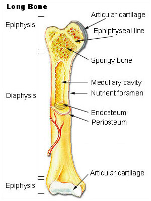
And recall anatomic structures in cross section. While it is not as hard as compact bone, spongy bone plays an important role of protecting the marrow where blood cells are produced. Shop the edit of floral dresses, dream jeans and fresh shoes now, and stay tuned for a lot more exciting topshop stuff to come. To the left is muscle tissue, and to the right is bone marrow. (area/long bone length 3) ∗ 10 8. The geometrical properties generated from the ct image included as follows: The compact bone is made up of osteon. Muscle attachments are visible along the outer surface. Would it be a good thing to show the epiphyseal plate? Internal structure of a human long bone, with a magnified cross section of the interior. Concentric layers of bone cells (osteocytes) and bone matrix surround the central canal. There are trabeculae in spongy bone which gives its sponge like appearance. The large dark spots are passages for blood vessels and nerves.
Shop the edit of floral dresses, dream jeans and fresh shoes now, and stay tuned for a lot more exciting topshop stuff to come. This is known as the periosteum. The upper (biting) surfaces of the tooth are at top, with the lower sections (bottom) embedded in the gums and jaw bone (not shown). The central tubular region of the bone, called the diaphysis, flares outward near the end to form the metaphysis, which contains a largely cancellous, or spongy, interior. Cross section of mandible at first molar region showing cortical and spongy bone basic concepts in osteogenesis.
Bone Marrow, Vertebral Body Cross-Section - Stock Image ... from media.sciencephoto.com Internal structure of a human long bone, with a magnified cross section of the interior. Bone is a dynamic biological tissue, composed of various metabolically active cells that are integrated into a rigid framework. In three dimensions an osteon is cylindrical in shape. The geometrical properties generated from the ct image included as follows: The cell line involved in osteogenesis consists of preosteoblasts, osteoblasts, osteocytes and bone. Body size standardization was done, using the following equations: Wing bones were sampled from the right side of skeletally table 1. Why is the marrow red?
Wing bones were sampled from the right side of skeletally table 1.
Shop the edit of floral dresses, dream jeans and fresh shoes now, and stay tuned for a lot more exciting topshop stuff to come. The central tubular region of the bone, called the diaphysis, flares outward near the end to form the metaphysis, which contains a largely cancellous, or spongy, interior. (area/long bone length 3) ∗ 10 8. Internal structure of a human long bone, with a magnified cross section of the interior. Now that you know what bones do, let's take a look at what they're made of and their anatomy. Internal structure of a human long bone, with a magnified cross section of the interior. Some of these resources are listed here. 100x first focus in the compact decalcified bone (cb) on the left part of the image, you can see small dots, which are. Internal structure of a human long bone. Sketch and label of a cross section of a long bone : Muscle attachments are visible along the outer surface. Eliminate sudden changes of direction and influx of one stream into another. In three dimensions an osteon is cylindrical in shape.
Bone is a dynamic biological tissue, composed of various metabolically active cells that are integrated into a rigid framework. Internal structure of a human long bone. Now that you know what bones do, let's take a look at what they're made of and their anatomy. Why is the marrow red? There are trabeculae in spongy bone which gives its sponge like appearance.
Animal Or Dinosaur Hip Bone/femoral Head? Alabama Origin ... from www.thefossilforum.com Cross section of mandible at first molar region showing cortical and spongy bone basic concepts in osteogenesis. This slide contained a cross section of a very small bone, and you are looking at the entire thickness of the shaft of the bone. 100x first focus in the compact decalcified bone (cb) on the left part of the image, you can see small dots, which are. The large dark spots are passages for blood vessels and nerves. An outer 'fibrous layer' containing mainly fibroblasts, and an inner 'cambium layer' containing progenitor cells. Would it be a good thing to show the epiphyseal plate? It consists of two layers; To the left is muscle tissue, and to the right is bone marrow.
Table 1 describes the bone markings, which are illustrated in (figure 4).
Now that you know what bones do, let's take a look at what they're made of and their anatomy. Bone markings the surface features of bones vary considerably, depending on the function and location in the body. Each bone in your body is made up of three main types of bone material: And recall anatomic structures in cross section. It consists of two layers; This slide contained a cross section of a very small bone, and you are looking at the entire thickness of the shaft of the bone. Body size standardization was done, using the following equations: Chapter 6 bones and skeletal tissues flashcards quizlet. Bone matrix and cells bone matrix osseous tissue is a connective tissue and like all connective tissues contains relatively few cells and large amounts of extracellular matrix. Compact bone, spongy bone, and bone marrow. The outlined area is a cross section of an osteon of compact bone. Start studying cross section of bone. I don't find it enhances the image.